
Fig 5.
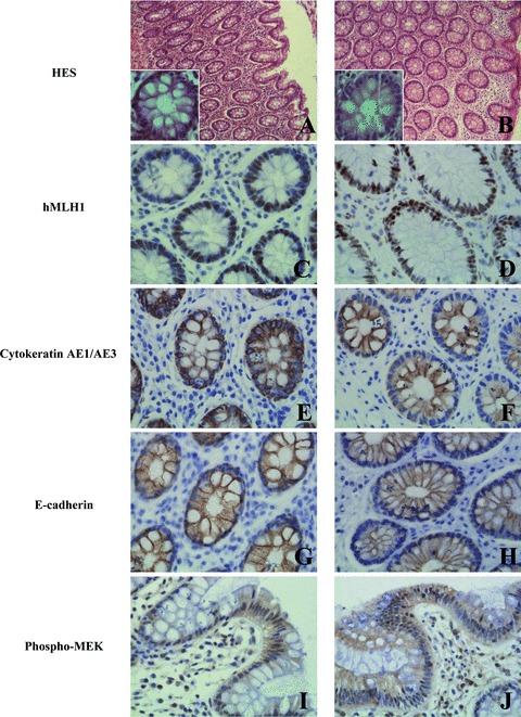
Morphology and immunohistochemical staining of hMLH1, cytokeratin AE1/AE3 and E-cadherin of normal colonic mucosa fixed with formalin and RCL2 on the left and right panels, respectively. HES stained sections (A, B); nuclear hMLH1 (C, D); cytoplasmic cytokeratin AE1/AE3 (E, F); membrane E-cadherin (G, H) and cytoplasmic and nuclear phospho-MEK (I, J) immunoreactivities are presented. Original magnification x 400. For panels A and B, original magnification x100, insets x400.