Fig 1.

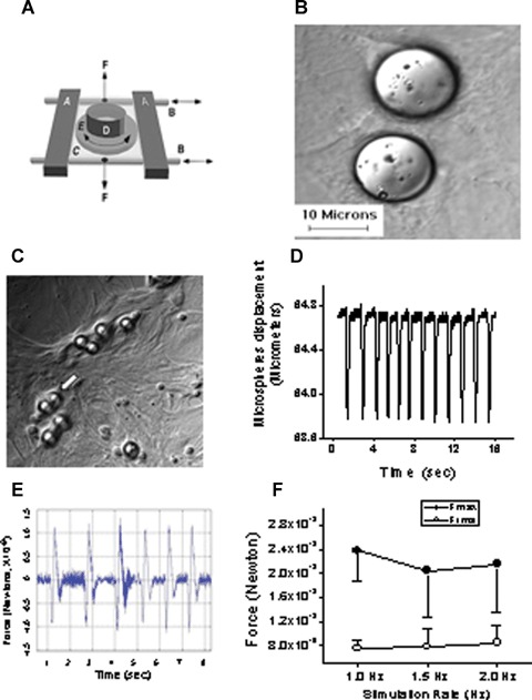
The experimental procedures for inducing mechanical load by means of cyclic stretch and glass microspheres in neonatal rat ventricular myocytes (NRVM) cultures. [A] Adopted from Ref. 14. Schematic presentation of the cyclic stretch apparatus designed and built by Dr. André Kléber (Department of Physiology, University of Bern, Bern, Switzerland). Horizontal metal bars (A) glide horizontally on stainless cylindrical axes (B) and support the transparent silicone membrane (C). A segment of sili-cone tubing (D) was glued on the silicone membrane to form the walls of the culture dish in which the neonatal rat myocytes were seeded and grown. Two clamps (F; positions indicated by vertical arrows) produced slight tension along the central axis of the stretch apparatus and thereby reduced transverse shrinking to <1%. [B] Microspheres are spread on the culture. [C] Cells adhere to the collagen coated microspheres, sometimes completely covering them (white arrow), to form a single contracting unit. [D] Representative displacement recording of a single microsphere, obtained by means of a video edge detector. [E] Calculated force/time relationship from the displacement graph shown in panel D. [F] The force generated at different stimulation rates (n = 4–6 myocytes). Fmax, maximal force; Frms, root mean squares of force over time.
