
Fig 1.
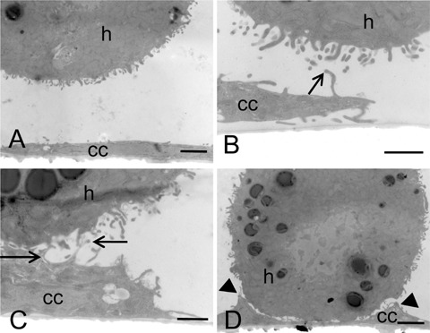
Electron micrographs of colon cancer cells and hepatocytes in coculture for 1 hr. CC531s cells were cultured on glass for 3 days and suspensions of hepatocytes were added. When hepatocytes were not in the vicinity, cancer cells (cc) were flattened with few small protrusions only and hepatocytes (h) were rounded (A). When hepatocytes were in close vicinity of cancer cells, cancer cells rapidly became bulky and formed protrusions in the direction of the hepatocytes (B). Contact between cancer cells and hepatocytes was established between cancer cell protrusions and microvilli of hepatocytes (arrows; B, C) and then stretches of parallel-running membranes were made (arrow heads; D). Bars = 2 pm (A, C) or 1 μmΊ (B, D).