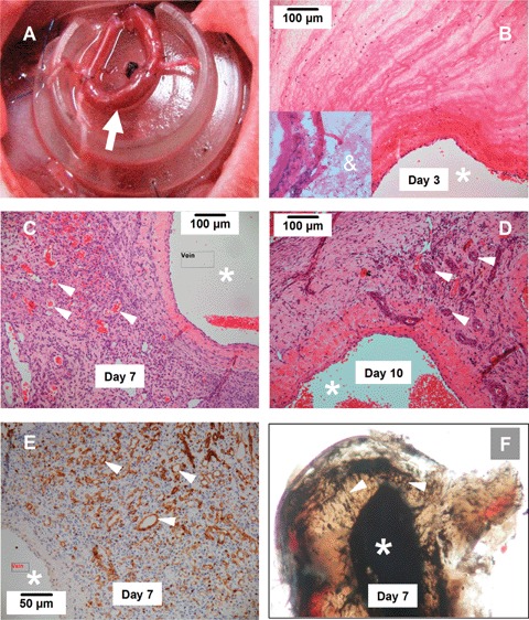
Fig 1.

Angiogenic response in tissue-engineering (TE) chamber (A). Arrow indicates the arterio-venous loop constructed by a vein graft anastomosed to the femoral artery and vein. (B-D) Histology of the tissue formed in the TE chamber 3, 7 and 10 days after chamber implantation (H&E staining). The lumen of the vein side of the parent loop was indicated by *. The new blood vessels are indicated by arrow heads. Note that vessels with a multi-layered wall composed of mural cells can be observed in 10-day specimen. The insert in B shows that 1 day after construction, the chamber contained only a fibrin mesh (&) and a few infiltrating cells. (E) New vessels defined by Griffonia Simplicifolia lectin staining (brown, arrow heads) at 7 days. (F) Ink perfusion of the AV loop at 7 days demonstrates new vessels (arrow heads) sprouting form the parent vessel lumen (femoral vein). At each time point, 5–6 animals were analysed.
