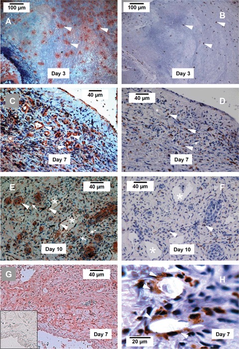
Fig 4.

Immunohistochemistry showing the expression of Nox2, Nox4 and the mono-cytes/macrophage marker ED-1. (A, C, E) Nox2 expression (red) in new blood vessels (arrow heads) and infiltrating leukocytes (arrows) in the chamber tissue at 3, 7 and 10 days. (B, D, F) Expression of ED-1 (brown) in infiltrating leukocytes (arrow heads) in the chamber tissue at 3, 7 and 10 days. (G) Expression of Nox4 (red colour) in the chamber tissue at 7 days. The insert shows a negative control section, in which non-specific IgG was used instead of the primary antibody. (H) We have observed that at day 7, some ED-1 + cells appeared to be incorporated in the new vessel (arrow heads). Such ED-1 + cells could not be observed in the vessels at day 10. Endothelial cells of the new vessel were ED-1 negative. * indicates the lumen of new blood vessels, n = 4 at each time point.
