
Fig 2.
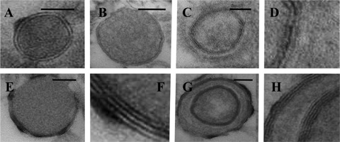
Typical appearance of matrix vesicles in the fibrous cap of atherosclerotic plaque (A-H). (A-B): Matrix vesicles surrounded by two electron-dense layers (lamellae) and filled with granular (A) or homogenous material (B) of medium electron density. (D-H): Matrix vesicles covered by multiple lamellae. (D) is a detail of (C). (F) is a detail of (E). (H) is a detail of (G). Transmission electron microscopy (TEM). Scale bars: 50 nm (A, B, C, EandG).