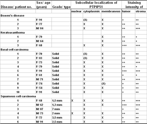
Fig 1.

Summary of PTPIP51 expression in keratinocyte carcinomas and precancerous lesions. Staining intensity was assessed as strong (+++), moderate (++), weak (+), not reactive (−) and the staining pattern was divided into three groups: Nuclear, cytoplasmic and membranous and was marked by an X or (X). X indicates that this particular PTPIP51 staining pattern was observed for the majority of cells (>75%) and X in brackets (X) means a staining pattern observed for the minority of cells (<25%). Basal cell carcinoma (BCCs) were graded according to the WHO classification, for squamous cell carcinoma (SCCs), the tumour thickness is given in mm.
