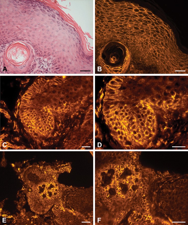
Fig 5.

H+E staining and PTPIP51-immunostaining of aSCC (A) H+E staining of epidermis next to SCC tissue with a horn pearl beneath. (B) PTPIP51-immunostaining of the same section displays a strong membranous and faint nuclear staining of keratinocytes. Cells lining the horn pearl presented a more intense signal. (C) Membranous PTPIP51-immunostaining of a SCC (D) High-power view of the same section. PTPIP51 staining reveals a punctiform localization-pattern. (E) Membranous PTPIP51 -immunostaining of a SCC. Some keratinocytes show an attenuated staining and some even lack PTPIP51 (F) Magnification of the same section. Bar: A, B: 50 μm, C–F: 20 μm.
