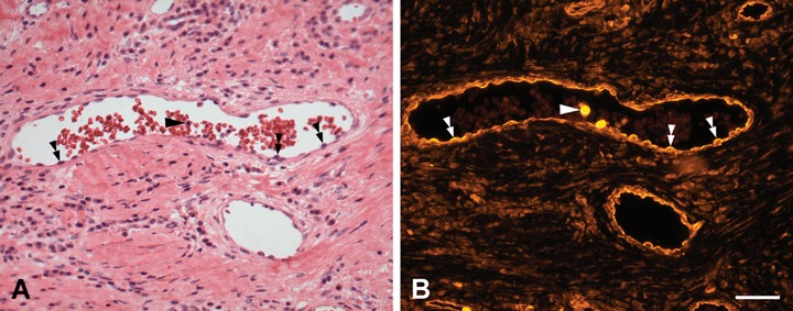
Fig 7.

H+E staining and PTPIP51-immunostaining of microvessels in the peritumoural stroma of a BCC (A) H+E staining (B) Immunostaining of the same section. PTPIP51 is expressed in endothelial cells (double arrowheads) and two neutrophils in the vessel lumen (arrow). Both immune cells show an intense signal of the PTPIP51-antibody. Bar: 50 μm.
