Fig 8.

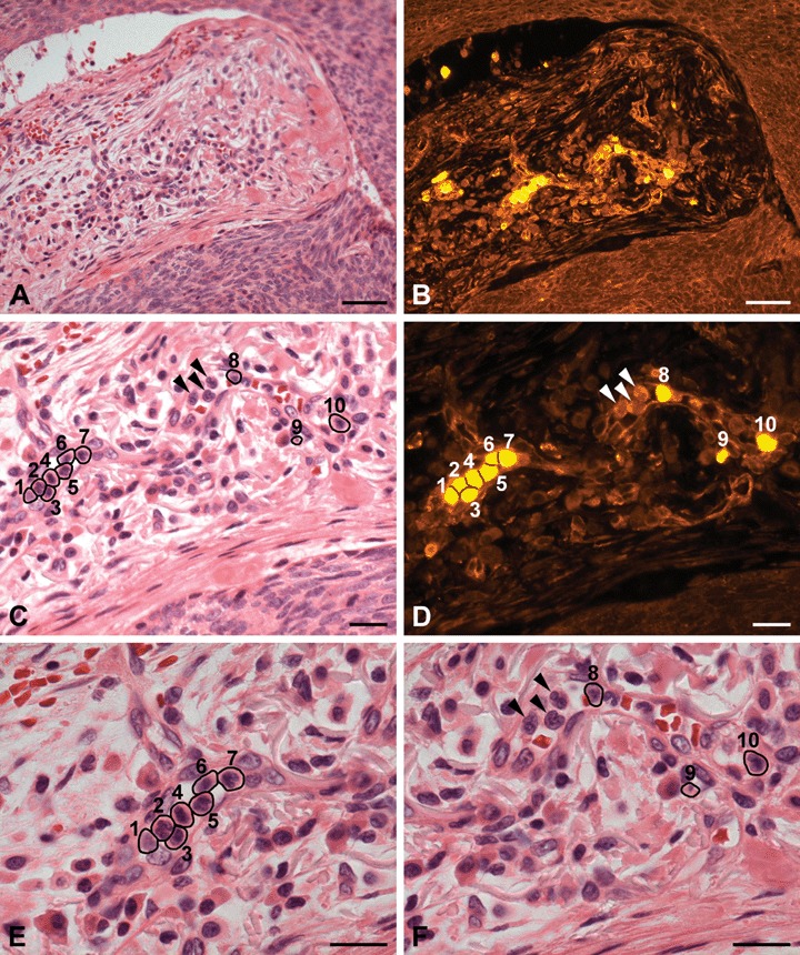
H+E staining and PTPIP51-immunostaining of a venous microvessel in the peritumoural tissue of a BCC. (A) H+E staining. Overview. (B) PTPIP51-immunostaining of the same section (C) H+E staining. Higher magnification of the same section. Multiple immune cells are located in the lumen of a capillary. The cells are numbered and marked by circlets. (D) PTPIP51-immunostaining of the same section. Not all immune cells (marked by arrowheads) are PTPIP51-positive. (E) and (F) provide a high-power view of (C) to identify PTPIP51-positive cells: 1–7: eosinophils and neutrophils; 8–10: neutrophils. As indicated by arrowheads, not all immune cells are PTPIP51-positive. Bar: A, B: 50 μm, C–F: 20 μm.
