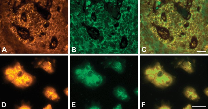
Fig 12.

Co-immunostaining of PTPIP51 and β-catenin in a human SCC (A–C) and HaCaT cells (D–F) (A) PTPIP51, (B) β-catenin-immunostaining of the same section, (C) Overlay. Co-localization is indicated by yellow colour. (D) PTPIP51. Note the predominant membranous localization. (E) β-catenin (F) Overlay. Co-localization is indicated by yellow colour. Bar: 20 μm.
