
Fig 8.
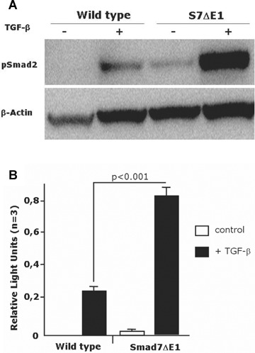
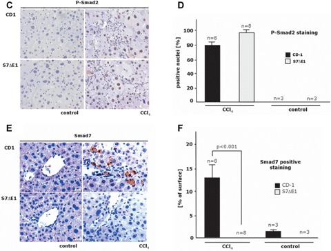
TGF^-induced signalling is enhanced in S7ΔE1 hepatocytes. (a) immunoblot analysis of Smad2 phosphorylation after 48 hrs of stimulation with 5 ng/ml TGF-β. A stronger band is observed in S7ΔE1 hepatocytes compared to wild-type cells. Notice that even under non-stimulated conditions S7ΔE1 cells yield a clear pSmad2 band that is not found with wild-type cells; (b) Smad3/4-luciferase reporter assays indicate that the stronger Smad phosphorylation observed in S7ΔE1 hepatocytes correlates with enhanced transcriptional activity of Smad complexes. Loss of Smad7 function leads to enhanced TGF-β signalling in a CCI4 model of liver damage (c-f). Representative photomicrographs of liver sections from CD-1 and S7ΔE1 strains treated or not (control) for 8 weeks with CCI4 are shown; (c) phospho-Smad2 immunostaining as a quantitative measurement for TGF-β signalling; (e) immunostaining for Smad7; (d, f) morphometric quantification of immunohistochemical stainings. Ten fields were selected randomly from each section of different groups (8 animals/group); about 200 cells were evaluated per observation field, immunopositive cells were counted and expressed as percentage of total cells investigated.