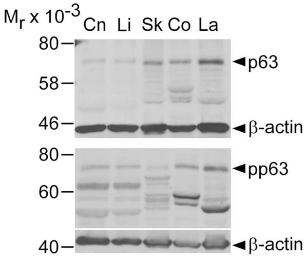
Figure 4.
Immunoblot analysis of equine epithelial tissues with anti-p63 and anti-phosphorylated p63 antibodies. Immunoblot analysis with antibodies against p63 (top panel) and pp63 (bottom panel) in equine corneal (Cn), limbal (Li), haired skin (Sk), coronary (Co) and mid-dorsal lamellar (La) tissues. Anti-β-actin antibody was used as a loading control. Numbers on the left indicate molecular mass standards (Mr × 10−3) while arrowheads on the right point to immunoreactive bands at the predicted molecular weight for p63 and pp63 (70-75 kDa).

