
Figure 1. Abluminal distribution of meningeal T cells and identification of Lyve-1 expressing vessels adjacent to the dural sinuses.
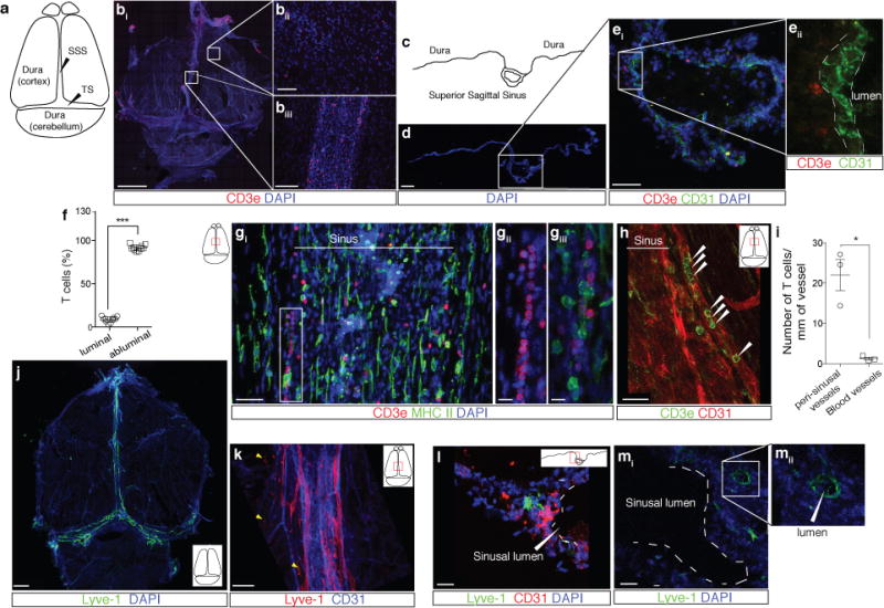
a. Schematic representation of the whole mount dissection of the dura mater. (SSS: Superior Sagittal Sinus; TS: Transverse Sinus) b. Representative images of CD3e labeling in whole mount meninges (scale bar = 2,000 μm). bii–biii. Higher magnification of the boxes highlighted in bi (scale bar = 90μm (bii) or 150 μm (biii)). c. Schematic representation of a coronal section of whole mount meninges. d. Representative image of a coronal section of whole mount meninges (scale bar = 200μm). e. Representative images of CD3e and CD31 immunolabeling in a coronal section of whole-mount meninges. Scale bar = 100 μm. eii. Higher magnification of the box highlighted in ei (scale bar = 30μm). f. Quantification of the percentage of sinusal T cells localized abluminally vs. luminally to the superior sagittal sinus (mean ± SEM; n = 18 fields analyzed from 3 independent animals; ***p=0.0008, Mann-Whitney test). g. Representative images of CD3e and MHCII-expressing cells around the superior sagittal sinus (meningeal cartoons here and elsewhere depict the location of the presented images; scale bar = 50 μm). gii. Higher magnification of the box highlighted in gi (scale bar = 10 μm). giii. High magnification of CD3 and MHCII-expressing cells (scale bar = 10 μm). h. Representative image of CD31 and CD3e labeling around the superior sagittal sinus (scale bar = 30 μm). i. Quantification of the number of T cells per mm of vessels in the perisinusal CD31+ vessels and in similar diameter meningeal blood vessels (mean ± SEM; n = 3 animals; *p=0.05; One-tailed Mann-Whitney test). j. Representative image of Lyve-1 labeling on whole-mount meninges (scale bar = 1,000 μm). k. Higher magnification of Lyve-1 expressing vessels (scale bar = 70 μm); arrowheads indicate Lyve-1+ macrophages. l. Representative images of CD31 and Lyve-1 labeling of a coronal section of the superior sagittal sinus (scale bar = 70 μm). mi. Higher magnification of a Lyve-1 positive vessel presenting a conduit-like structure (scale bar = 50 μm). mii. Higher magnification of the Lyve-1+ vessel presented in panel mi; arrowhead points to the lumen of the vessel.