
Figure 3. Functional characterization of meningeal lymphatic vessels.
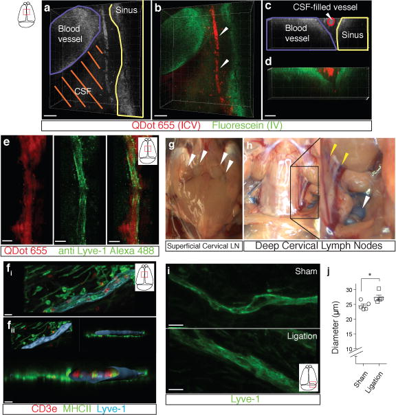
a–d. Representative z-stacks of the superior sagittal sinus of adult mice injected intravenously (i.v.) with fluorescein and intracerebroventricularly (i.c.v.) with QDot655 (n = 3 mice). a–b Low magnification images are presented showing fluorescein labeling in a meningeal blood vessel and in the superior sagittal sinus. In contrast, QDot655 labeling is prominent in the perisinusal vessel. c–d. Coronal section of the z-stack presented in panels a and b (scale bar = 20μm c–d). e. Representative z-stack of CSF-filled vessel from a mouse injected i.c.v. with both QDot655 and alexa488-conjugated anti-Lyve-1 antibody (n =3 mice; scale bar = 30μm). fi. Representative image of immunolabeling for CD3e and MHCII along with Lyve-1 in the meninges (scale bar = 15μm). fii. Representative image of a 3D reconstruction of the meningeal lymphatic vessels showing the luminal localization of the CD3e and MHCII-expressing cells (scale bar = 20 μm). g–h. Adult mice were injected i.c.v. with 5μl of 10% Evans blue. Superficial cervical lymph nodes (g) and deep cervical lymph nodes (h) were analyzed 30 min after injection (n = 5 mice); white arrowheads indicate the lymph nodes (g–h); yellow arrowheads indicate the Evans blue filled vessels arising near the internal jugular vein into the deep cervical lymph nodes (h). i–j. The collecting vessels draining into the deep cervical lymph nodes (yellow arrowheads in h) were ligated or sham-operated. Eight hours after the ligation, the meninges were collected and immunolabeled for Lyve-1. Representative images of immunolabeling for Lyve-1 in the transverse sinus of ligated and sham-operated mice (i; scale bar = 30 μm). Dot plots represent measurement of the meningeal lymphatic vessel diameters (j; mean ± SEM; n = 5 mice each group from 2 independent experiments; *p=0.031; Mann-Whitney test).