
Extended Figure 4. Pilot identification of lymphatic vessels in human dura.
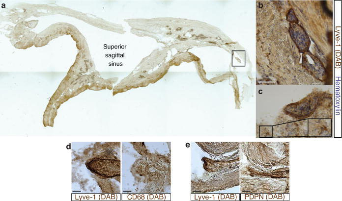
a. Representative image of a formalin-fixed coronal section of human superior sagittal sinus. b–c. Representative images of Lyve-1 staining on coronal section of human superior sagittal sinus (scale bar = 100 μm). The box in c highlights the presence of Lyve-1 expressing macrophages in human meninges, as seen in mice. d. Representative images of Lyve-1 and CD68 staining of coronal sections of human superior sagittal sinus. Note the absence of CD68 positivity on Lyve-1 positive structures (scale bar = 50 μm). e. Representative images of podoplanin and Lyve-1 staining of coronal sections of human superior sagittal sinus (scale bar = 50 μm).