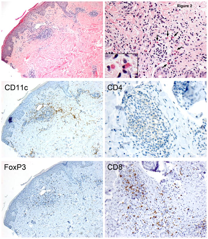
Figure 2. Vaccine-induced delayed-type hypersensitivity reactions to irradiated autologous sarcoma cells.
A representative analysis of a skin biopsy obtained 2–3 days after the second injection of irradiated autologous sarcoma cells. Shown are the H&E staining and immunohistochemistry for CD1a, CD11c, CD4, FoxP3, and CD8 expressing dendritic cells and T cells (400×). A prominent interface perivascular infiltrate is seen with eosinophils.

