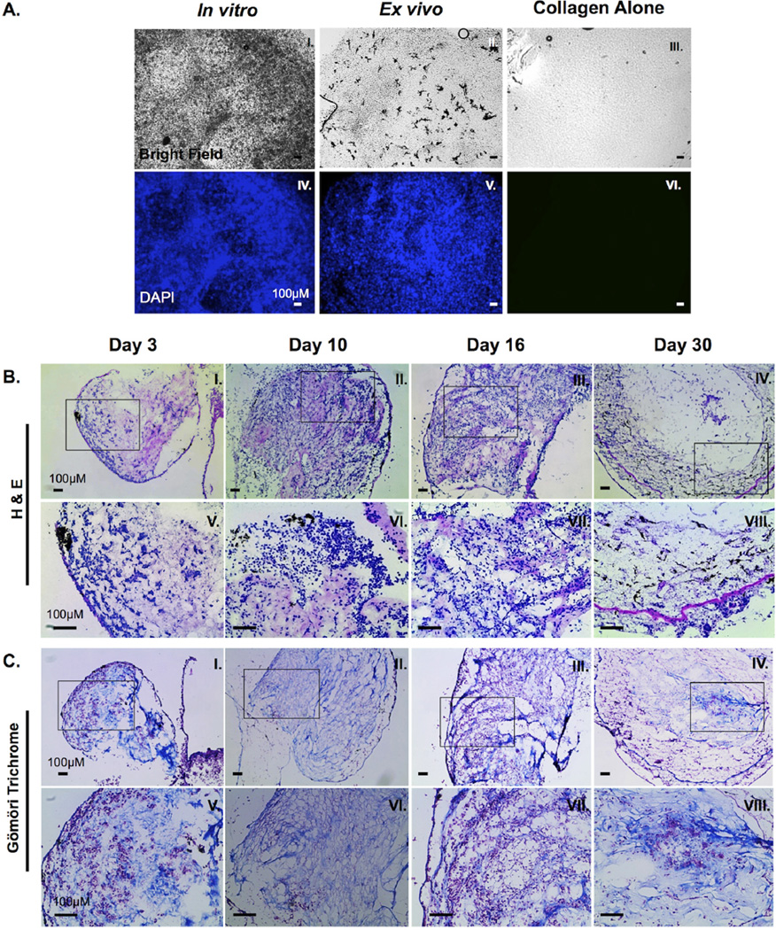
Figure 2. Characterization of 15/0 tumor cells within the semi-solid tumor graft.
(A) Semi-solid tumors containing 500,000 15/0 tumor cells were either grown for 9 days in vitro (I and IV) or grafted subcutaneously in vivo in LG-15 tadpoles (II and V). Control collagen mass without tumor cells was maintained in vitro (III and VI). Semi-solid tumors were then stained with 0.1ug/ml DAPI (II, IV, and VI) and cells visualized using a Leica DMIRB inverted fluorescence microscope. (B) H&E staining of cryosections of tumor collagen grafts 3, 10, 16 and 30 days post transplantation (I–IV). Magnified images of boxed section from above panel (V–VIII). Slides were imaged using an Axiovert 200 inverted microscope. (C) Gömöri trichrome staining of cryosections of tumor collagen grafts 3, 10, 16 and 30 days post transplantation (I–IV). Magnified images of boxed section from above panel (V–VIII). Slides were visualized using an Axiovert 200 inverted microscope.

