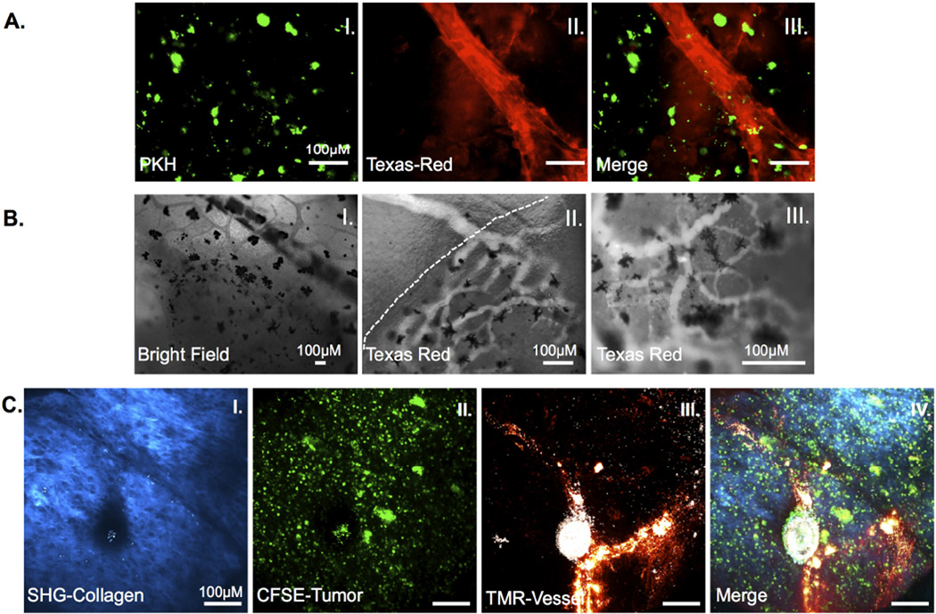
Figure 3. Neo-angiogenesis in 15/0 collagen grafts.
LG-6 or LG-15 tadpoles were grafted with tumor grafts containing 500,000 15/0 tumor cells and animals were intracardiacally injected with either Texas red 70KDa or TMR 2000KDa dextran and imaged. (A) Ex vivo images of vascularized grafts from LG-15 tadpoles at day 7 post transplantation. PKH-labeled 15/0 cells (I) with Texas red-dextran filled vasculature (II). Vascularized tumors were imaged ex vivo using an Olympus BX40 conventional fluorescence microscope. (B) Intravital imaging of vasculature in LG-6 15/0 collagen grafted animals. Bright field image of vasculature entering tumor mass (I). Texas-red dextral filled vasculature entering semi-solid tumor (dashed line indicates tumor boundary) (II). Texas-red dextral filled vasculature within tumor mass (III). Black cells are melanophores within the tumor graft. Animals were imaged using a Nikon TE2000-U epifluoresence microscope. (C) TMR loaded vasculature within 15/0-collagen tumors in LG-6 tadpoles showing SHG signal (I), CFSE labeled tumor (II) and TMR loaded vessels (III) from 15/0-collagen tumor bearing animals. Animals were imaged using an Olympus two-photon laser-scanning microscope. Images from representative animals shown.

