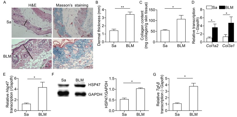
Figure 3.

Expression of HSP47 increased in the skin lesions of mice treated with BLM. (A) Hematoxylin and eosin (H&E) and Trichromestaining for the skin tissues of normal control mice and BLM-treated mice, Original magnifications were 200×, and the length of bars in the figures are 100 μm; (B) Dermal thickness was calculated at 10× microscopic magnification by measuring the distance between the dermal-epidermal junction and the derma-subcutaneous fat junction (μm) (as indicated by H&E staining of in Fig. 1A) in five randomly selected fields for each skin section. (C) Deposition of collagen in the skin was examined by Sircol assay. RT-PCR was used to analyze the expression of collagen (D), Hsp47 (E) and Tgf-β (G) in the skin of mice, and the mRNA levels were calculated using a relative ratio to Gapdh. (F) Western blots for the protein level of HSP47 in mice skin, and densitometric data of Western blot for HSP47 was shown in the column chart. Bars show the means ± SEM results from 6 mice in each group. *P < 0.05, **P < 0.01 versus saline treated group (Sa). Each group n = 6
