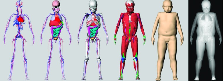
FIG. 5.
The different layers of a 9-yr-old female phantom (85th percentile in body mass and 11th percentile in height). From left to right: vasculature; organs; skeleton; muscle, tendons, and ligaments; and skin surface. A simulated x ray projection is shown to the far right demonstrating the use of the phantom for imaging simulation.

