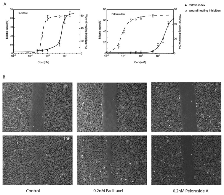
Figure 2. Drug effects on wound healing.
(A) Dose response curves for the inhibition of mitosis (measured as the mitotic index; i.e., the percentage of accumulated mitotic cells after 24 h) and the inhibition of wound healing after 10 h are shown in panel A for paclitaxel or peloruside A. For wound healing, a scratch wound was introduced into near confluent layers of HUVEC and the rate of wound closure was measured in the presence or absence of paclitaxel or peloruside A at the specified concentrations. (B) Panel B shows examples of the monolayers at 1 hour or 10 hours after introduction of the scratch under control conditions or at 0.2 nM of the specified drug. Scale bar in panel A is 100 μm.

