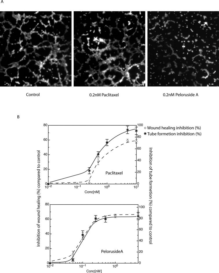
Figure 4. Comparison of the abilities of paclitaxel and peloruside A to inhibit capillary tube formation.
Endothelial cells (HUVEC) were plated at equal densities onto Matrigel in the presence and absence of the specified concentrations of paclitaxel or peloruside A. Cells were then examined after 5 h for the ability to form tubes. (A) Images of capillary tube formation at a single concentration of each drug are shown in panel A. (B) Dose response curves showing the inhibition of capillary tube formation as compared to wound healing is shown in panel B.

