
Fig. 1.
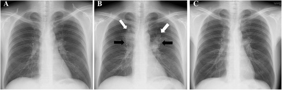
Serial changes on chest radiograph. a Chest radiograph taken at the time of starting combined antiretroviral therapy (cART), showing almost normal findings. b Chest radiograph taken five months after starting cART, showing infiltrates in the bilateral upper lung fields (white arrow) and bilateral hilar lymphadenopathy (black arrow). c Chest radiograph taken one year after starting anti-mycobacterial chemotherapy, showing slight hilar lymphadenopathy