
Fig. 1.
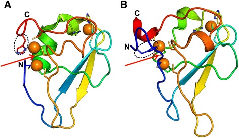
Structure of the UBR-box (a) UBR-box from human UBR1 (PDB identifier 3NY1_A) (b) UBR-box from S. cerevisiae UBR1 (PDB identifier 3NIH_A). The structures have been colored from the N- to C-terminal in a gradient of blue to red. The zinc-chelating residue which displays circular permutation in S. cerevisiae UBR-box with respect to the human UBR-box is marked with a dotted circle. The shared metal-chelating residue is indicated by a red arrow