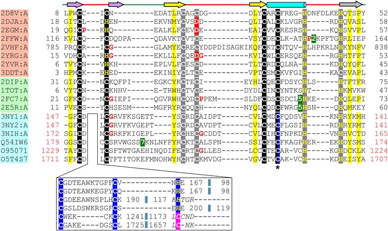
Fig. 3.

Structure based multiple sequence alignment of B-box, ZZ domain and UBR-box. PDB/UniProt identifier, start and end aminoacid numbers are indicated for each sequence. Identifiers of the representative sequences of the B-box are highlighted in peach, ZZ domain in light green and UBR-box in light blue. Secondary structure diagram is depicted above the alignment. The zinc-binding residues (Cys/His) of the first zinc binding site of the binuclear treble clef have been highlighted in black, those of the second zinc binding site in grey, those of the third zinc binding site (for the UBR-box) in dark blue and other aminoacids at equivalent position in red. Residues that may potentially serve as metal-chelating ligands are highlighted in pink. The shared ligand in the UBR-box is indicated by an asterisk (*) and highlighted in black and blue. The sequence region in between the circularly permuted zinc-knuckle of the UBR-box, which is not present in the B-box and the ZZ domain, is shown in a separate box under the alignment of the common binuclear RING-like regions. Regions of circular permutation are separated by a small blue colored box and sequence numbers of the regions around the circular permutation are colored in red. Regions where the structures are not superimposable are shown in italics. Small aminoacids (Gly, Pro) in the vicinity of the zinc-binding ligands are colored red. Uncharged residues (all aminoacids except Asp, Glu, Lys and Arg) in mostly hydrophobic sites are highlighted yellow. Long insertions are not shown and the number of omitted residues is boxed in green
