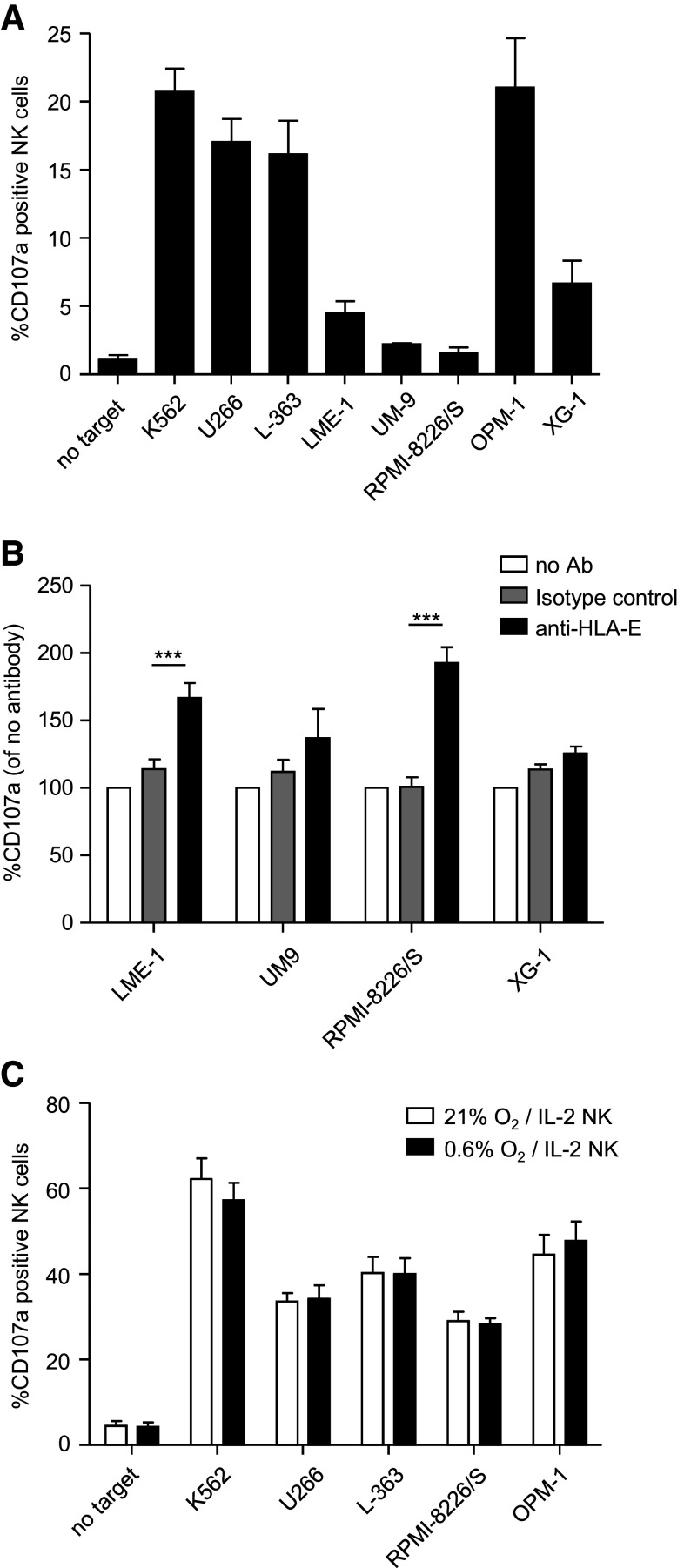
Fig. 5.
HLA-E can abrogate the anti-myeloma response of NKG2A+ NK cells. a The tumor cell lines were co-cultured at 21 % O2 with freshly isolated peripheral blood-derived NK cells for 12 h, in the same assay as in Fig. 4 a/c. NK cells expressing only NKG2A as an inhibitory receptor were analyzed downstream for CD107a expression. Graph shows mean percentage and SEM CD107a+ of NKG2A+ KIR− NK cells. b LME-1, UM-9, RPMI-8226/S and XG-1 were co-cultured with non-activated NK cells for 12 h at 21 % O2, in the presence of an HLA-E-blocking antibody (black bars), an IgG1 isotype control (gray bars) or without antibody (white bars). Graph shows mean normalized percentage of CD107a+ cells of NKG2A+ KIR− NK cells (% with Ab divided by % without antibody). c Myeloma cell lines were pre-incubated for 6 h at 21 % or 0.6 % O2. NK cells were activated with 1000 U/ml IL-2 at 21 % O2. Thereafter, they were co-cultured with K562, U266, L363, RPMI-8226/S and OPM-1 for 10 h at 21 % O2 or at 0.6 % O2, and CD107a expression on KIR-NKG2A+ NK cells was analyzed as described in Fig. 4. Graph shows mean percentage and SEM CD107a+ of NKG2A+ KIR− NK cells. All experiments were performed in duplicate and with n = 5 donors. ***p < 0.001

