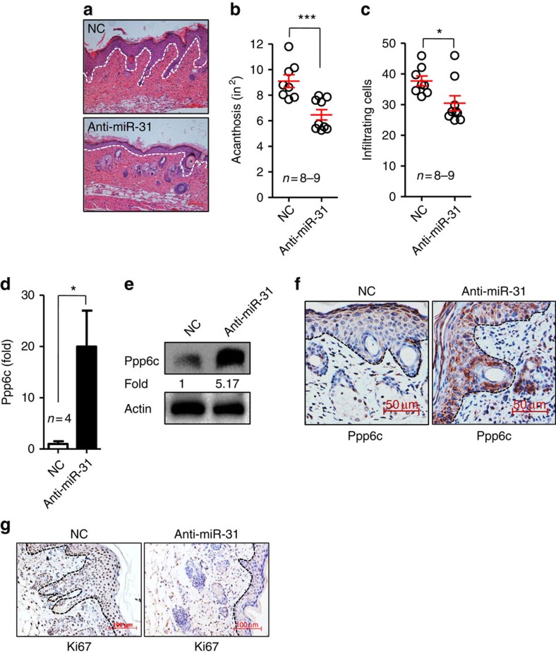
Figure 7. Administration of antagomir-31 decreases epidermal hyperplasia and dermal cellular infiltration induced by IMQ.
Mice were injected subcutaneously with an irrelevant antagomir (NC) or an antagomir to miR-31 (anti-miR-31). The first injection was administered 3 days before the application of IMQ and thereafter was performed every other day until the end of the experiment. (a) H&E staining of the back skin derived from mice injected with NC (upper panel) or anti-miR-31 (lower panel). Scale bar, 100 μm. (b,c) Acanthosis and dermal cellular infiltrates were quantitated for mice treated with NC or anti-miR-31. (d,e) Ppp6c mRNA and protein levels in NC- or anti-miR-31-treated mice. (f,g) Immunohistochemical staining of ppp6c or Ki67 in skin sections derived from NC- or anti-miR-31-treated mice after induction of skin phenotype by IMQ (n=8–9). Scale bar, 50 μm (f) or 100 μm (g). For all measurements (c), the median number of specifically stained dermal nucleated cells was counted in three high-power fields per section. Results (d) are presented as the ratio of mRNA to the β-actin, relative to that in NC-treated mice. *P<0.05, ***P<0.001, two-tailed Student's t-test. Data (a–g) are representative of at least two independent experiments with four to nine samples per group in each (mean and s.e.m.).

