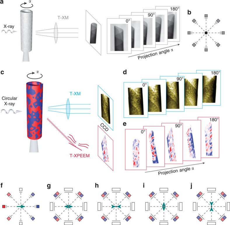
Figure 1. Comparison of the conceptual difference between conventional scalar tomography and MXT.
(a) Light is attenuated when penetrating an object, for example, rolled-up tubular magnetic nanomembrane, according to its local atomic mass distribution. (b) The spatial distribution in 3D space is obtained by applying tomographic reconstruction algorithms relying on the angle invariance of the scalar properties. (c) The magnetization component along the beam propagation direction can be visualized utilizing XMCD with soft X-ray microscopies. (d,e) exemplarily show the XMCD contrast patterns of radially and in-plane magnetized tubular architectures recorded with a TXM and a T-XPEEM, respectively. (f) The XMCD signal shows an angular dependence that even reverses its sign for space-inverted X-ray propagation (red to blue). The corresponding underdetermined system of projections leads to an ambiguity of possible reconstruction when considering arrangements of two or more macrospins (g–j) that may only be released by locating the magnetic material, determining the preferential magnetization orientation and analysing their evolution with varying projection angle. Determining all magnetization vector components requires to record projections around several rotation axes. The present case of tubular surfaces with either radial or in-plane magnetization allows for retrieving the magnetization textures from one set of projections taken around a rotation axis that coincides with the symmetry axis.

