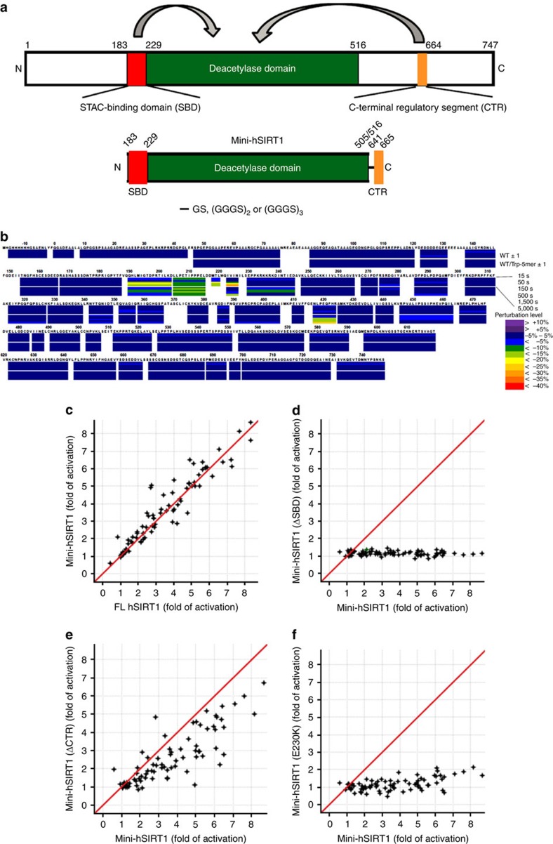
Figure 1. Mini-hSIRT1 construct design and characterization.
(a) Schematic diagram of human full-length hSIRT1 and Mini-hSIRT1 constructs. The N-terminal SBD, the central catalytic domain and the CTR are highlighted in red, green and orange. (b) Heat map of the HDX-MS perturbation of binding of 1 to hSIRT1 in the absence or presence of Ac-p53(W5) (Trp-5mer) at six different time points (15–5000, s). (c) Pivot plot of the activation by a chemically diverse STAC set using the Ac-p53(W5) substrate for mini-hSIRT1 versus full-length hSIRT1, as measured by OAcADPR assay. The red line represents y=x correlation. (d) Pivot plot of the STAC activation of mini-hSIRT1(ΔSBD) versus mini-hSIRT1. (e) Pivot plot the STAC activation of mini-hSIRT1(ΔCTR) versus mini-hSIRT1. (f) Pivot plot of the STAC activation of mini-hSIRT1(E230K) versus mini-hSIRT1.

