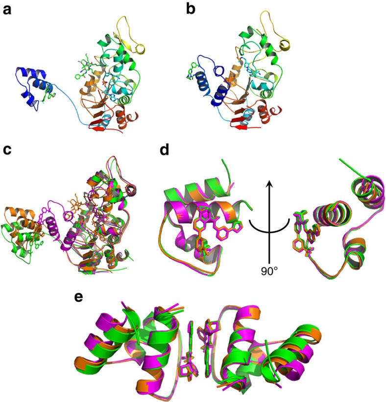
Figure 3. Structures of Mini-hSIRT1-STAC/ligand complex.
(a) Structure of mini-hSIRT1/1/Ac-p53 7-mer/CarbaNAD quaternary complex. The STAC 1 and Ac-p53 7-mer are shown in green, red and blue for carbon, oxygen and nitrogen atoms. The CarbaNAD is shown in cyan, red, blue and orange for carbon, oxygen, nitrogen and phosphate atoms. The protein ribbon is rainbow-colored from blue at the N-terminus to red at the C-terminus. (b) Structure of mini-hSIRT1/1/2 complex. The mini-hSIRT1 shown in this complex is hSIRT1(183–516)-GS-CTR as the same complex containing hSIRT1(183–505)-(GGGS)2-CTR diffracts to 3.5 Å even though the structures are almost identical. The STAC 1 is shown in green, red and blue for carbon, oxygen and nitrogen atoms. The Inhibitor 2 is shown in cyan, red, blue and yellow for carbon, oxygen, nitrogen and sulfur atoms. The protein ribbon is rainbow-colored from blue at the N-terminus to red at the C-terminus. (c) Structural comparison of mini-hSIRT1/1 complex (green), mini-hSIRT1/1/Ac-p53 7-mer/CarbaNAD quaternary complex (orange) and mini-hSIRT1/1/2 complex (magenta). (d) Superimposition of the SBD domains from mini-hSIRT1/1 complex (green), mini-hSIRT1/1/Ac-p53 7-mer/CarbaNAD quaternary complex (orange) and mini-hSIRT1/1/2 complex (magenta). (e) Comparison of the STAC-mediated dimer interface of mini-hSIRT1/1 complex (green), mini-hSIRT1/1/Ac-p53 7-mer/CarbaNAD quaternary complex (orange) and mini-hSIRT1/1/2 complex (magenta).

