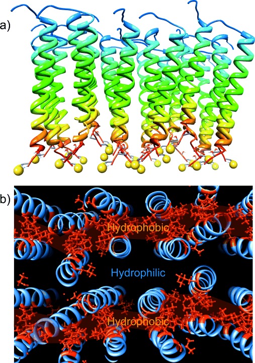
Figure 6.

Representative snapshots of MD simulations of a) side view and b) top view of a monolayer assembled on Au from 24 individual BASE-C peptides. The yellow spheres are sulfur moieties associated with the cysteine residue. The gold surface has been removed for clarity. Part (b) also plots the organization of hydrophobic leucine residues (highlighted in red) within the peptide monolayer.
