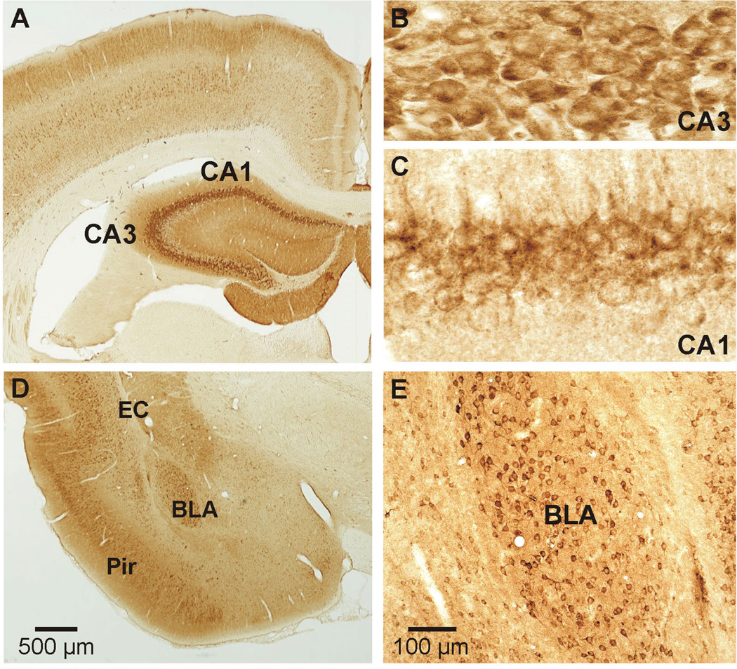
Figure 4.
Localization of AMPA receptors in a coronal section of the rat brain showing distribution in areas relevant to epilepsies. Immunostaining was carried out with the immunoperoxidase method, using a specific antibody to the AMPA receptor GluA2 subunit. (A), Low-power view of hippocampus and surrounding neocortex. There is dense staining of pyramidal cells in the CA3 and CA1 subfields. (B) and (C), Higher-power view of stained neuronal cell bodies and processes in CA3 (B) and CA1 (C). (D), Low-power view of the anterior portion of the amygdala showing the external capsule (EC), basolateral amygdala (BLA), and piriform cortex (Pir). (E), Higher-power view of the BLA showing prominent staining of neuronal cell bodies. Scale in (D) also applies to (A). Reproduced from (35), with permission of John Wiley & Sons.

