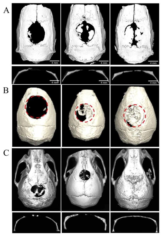
Figure 4.
Bone regeneration through different miRNA treatment. (A) Coronal and sagittal views of the harvested skulls by micro-CT at week 8 post-implantation (left: PGS scaffold, middle: anti-miR-negative/BMSCs/PGS, right: anti-miR-31/BMSCs/PGS). Adapted and reprinted with permission from Ref. [50]. (B) Micro-CT of nude mice at week 12 after surgery. (left: hASCs/scaffolds, middle: Bac-FLPo/Bac-FCBW/hASCs/scaffolds, right: Bac-FLPo/Bac-FCBW/Bac-miR-148b/hASCs/scaffolds). Adapted and reprinted with permission from Ref. [51]. (C) Micro-CT of live mice at week 12 after surgery. (left: BMSCs/hydrogel, middle: miR-negative/BMSCs/hydrogel, right: miR-26a/BMSCs/hydrogel). Adapted and reprinted with permission from Ref. [52].

