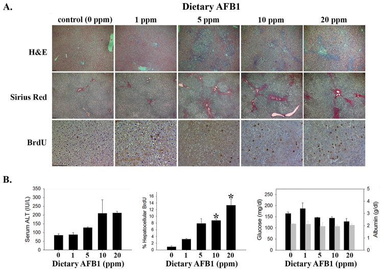
Figure 2. Dietary AFB1 Induced Liver Injury in Rats.
(A) AFB1-exposed rats demonstrate dose-dependent increases in bile duct hyperplasia (by H&E staining, image photographed at 50x magnification), fibrosis (by Sirius Red, 50x), and hepatocellular proliferation (by immunohistochemical detection of BrdU incorporation, 200x; 100 micron bar in lower left corner of left-most image in each series). (B) Quantification of serum ALT (left panel, ANOVA p=0.04); hepatocellular BrdU (middle panel, ANOVA p=0.02; *p<0.05 vs. control); serum glucose (black bars) and serum albumin (grey bars; right panel).

