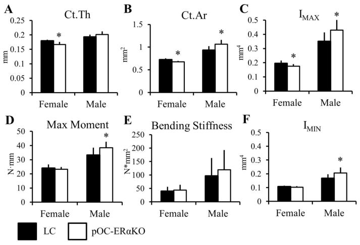
Figure 2.
Femoral midshaft bone morphology and strength were differentially affected in 12-week-old pOC-ERαKO females and males compared to LC. Female pOC-ERαKO mice had (A) decreased Ct. Th, (B) decreased Ct.Ar, and (C) decreased IMAX compared to LC. (D,E) Maximum moment and bending stiffness were not different between genotypes in females from 3-point bending mechanical tests. Male pOC-ERαKO mice exhibited an opposite bone phenotype compared to LC than that found in females. (B) Ct.Ar, (C) IMAX, and (F) IMIN were all increased in male pOC-ERαKO mice, which resulted in (D) increased maximum moment in 3-point bending tests.
Ct.Ar, cortical area; Ct.Th, cortical thickness; IMAX and IMIN, maximum and minimum moments of inertia. Data are mean ± SD, n=8–12 per group. *pOC-ERαKO different from LC, p<0.05 by one-way ANOVA for each sex.

