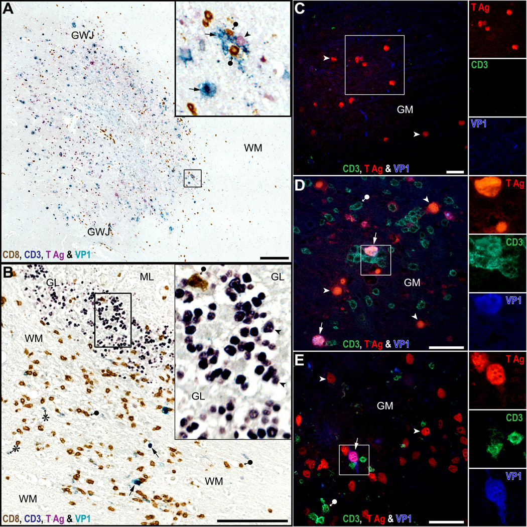
Figure 3.
CD8-positive T-cells aggregate mostly around cells with intact nuclei, expressing JC virus (JCV) T antigen (T Ag) and VP1. (A, B) Quadruple immunostaining for CD8 (brown), CD3 (grey blue), T Ag (purple) and VP1 (cyan-blue) done on an HIV-seronegative PML patient with chronic lymphocytic leukemia (A) and an HIV-seropositive PML patient (B). In PML lesions of the cerebral gray-white junction (GWJ) and gray matter (GM) (A, inset), where JCV-infected cells expressing VP1 are present (cyan-blue, arrows), there are numerous parenchymal CD8-positive T-cells (brown, black circle arrow). They are located mostly at the leading edge of infection where newly infected cells have still intact nuclei and express T Ag alone (purple, arrowhead) or T Ag and VP1 (cyan-blue, arrows). In JCV-infected cerebellum (B), only rare CD8-positive T-cells (brown, filled circle-arrow) colocalize in the GL with hundreds of JCV-infected cells expressing T Ag (inset, purple, arrowheads), while more numerous CD8-positive T-cells and some CD4-positive T-cells (grey-blue, asterisk) colocalize in the contiguous WM with a few JCV-infected cells expressing VP1 (cyan-blue, arrow). (C–E) Triple immunofluorescence (IFA) staining for CD3, T Ag and VP1 done in the cerebrum of an HIV-seronegative PML patient (C), an HIV seropositive PML patient (D) and a JCV encephalopathy (JCVE) patient (E). The IFA immunostains confirm that T-cells are absent in an area with T Ag only (C), whereas T-cells are mostly present in areas where JCV-infected cells have intact nuclei with co-expression of T Ag and VP1 (pink, arrows in squares in D and E). Separate color channels are shown in insets. Scale bars: A, B, 250 µm; C-E, 100 µm.

