
Figure 4.
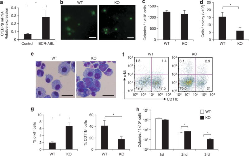
BCR–ABL-mediated in vitro colony formation in the absence of C/EBPβ. (a) C/EBPβ mRNA levels in c-kit+ Sca-1+ Lin− cells from WT bone marrow cells transduced with a control MIG vector or a MIG-BCR–ABL vector (n=3 each, P<0.01). (b) Colonies formed by BCR–ABL-transduced C/EBPβ KO bone marrow cells and WT bone marrow cells after culture for 7 days in cytokine-free methylcellulose (scale bar, 1 mm). Colony numbers (c), cell numbers per colony (d), and Wright Giemsa staining (scale bar, 20 mm; original magnification, ×400) (e) of the colony-forming cells are shown. Error bars indicate s.d. from triplicate cultures. Results are representative of three independent experiments. *P<0.01. (f) Flow cytometric analysis of the cells forming primary colonies by day 7. Numbers in each quadrant indicate the percentage of live cells. (g) Frequency of c-kit+ cells and CD11b+ cells in the cells forming primary colonies by day 7. Results are representative of two independent experiments. *P<0.05. (h) Serial colony-replating of 1×10⁴ BCR–ABL-transduced C/EBPβ WT and KO bone marrow cells. The colonies were counted and collected on day 10 (second) or 14 (third), respectively. Error bars indicate s.d. from triplicate cultures. Results are representative of three independent experiments. *P<0.01.