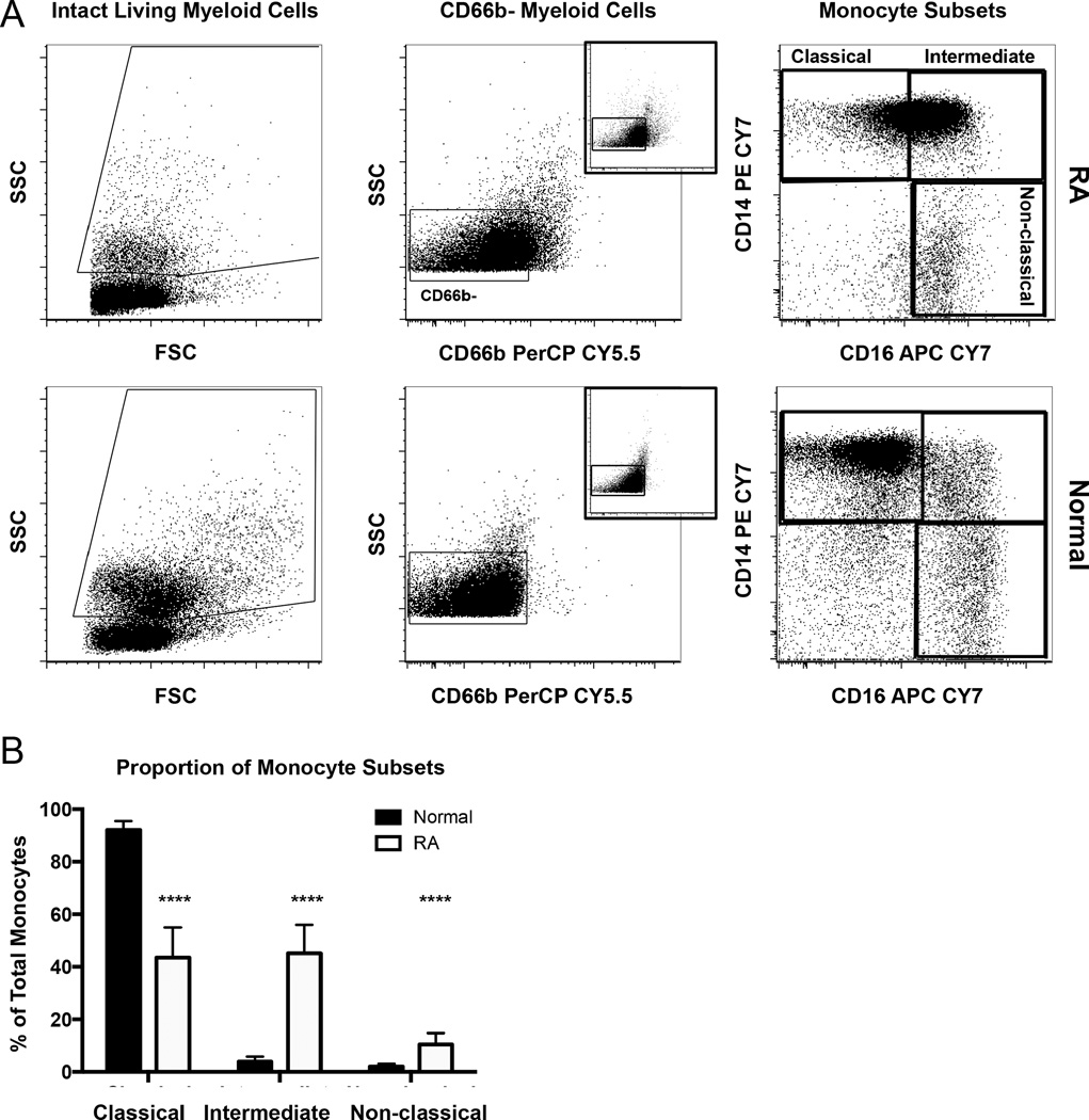
Figure 4.
The circulating monocyte pool is skewed toward intermediate and nonclassical monocytes in RA. A. PBMC from patients with RA and normal donors were stained for flow cytometry to detect CD66b, CD14, and CD16. Myeloid cells were gated (left panels) as in Figure 1. Expression of CD66b versus SSC was plotted, and monocytes were gated based on low side scatter and absence of CD66b (middle panels). Co-staining with CD14 and CD16 was used to identify 3 subsets of monocytes (right panels). B. The percentage of each subset within the total monocyte pool in healthy donors (n = 16) and patients with RA (n = 15). **** p ≤ 0.0001. RA: rheumatoid arthritis; PBMC: peripheral blood mononuclear cells; SSC: side light scatter.

