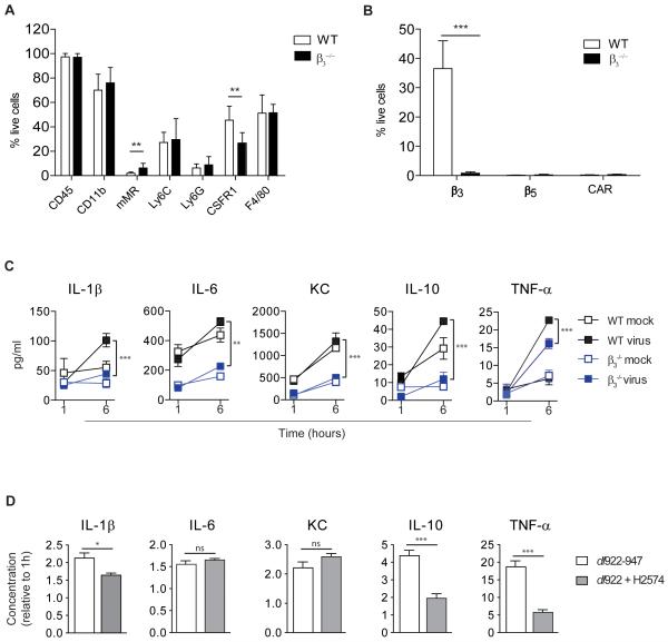
Fig.5. Cytokine release by murine peritoneal macrophages is β3 integrin-dependent.
Peritoneal cells from female WT and β3−/− mice were pooled for each genotype and assessed by multichannel FACS. Experiments were carried out in triplicate and data are presented as mean±SD (n=4-24).
A. Expression of macrophage cell surface markers (**p<0.01).
B. Expression of the adenoviral receptors, CAR, β3 and β5 (***p<0.001).
C. Murine peritoneal cells were treated ex vivo with dl922-947 (10,000vp/cell) or vehicle. Cytokine protein in supernatants was quantified at 1 and 6 hours. Change over time was compared in WT and β3−/− peritoneal cells. (**p<0.01, ***p<0.001). KC=keratinocyte chemoattractant
D. Murine peritoneal cells were pooled and treated ex vivo with dl922-947 (10,000vp/cell) alone or in combination with the β3 inhibitor, H2574 (20μM). Cytokine protein is shown at 6 hours relative to 1 hour (*p<0.05, ***p<0.001, ns=non-significant). KC=keratinocyte chemoattractant

