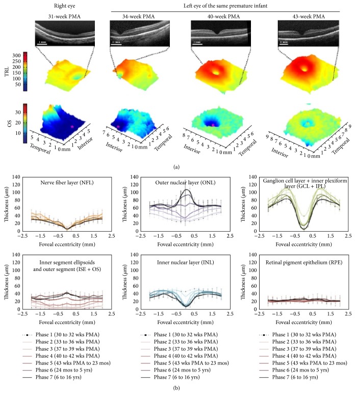
Figure 4.
3D map of retinal layers and their dynamic changes with age in a neonate. The lower portion of the image has a segment on inner segment ellipsoid (photo courtesy: for the top of the figure (color maps), taken from Figure 2(a), Page 2320 of [16]; for the bottom portion of the figure (graphs), taken from Figure 2, Page 782. of [17]).

