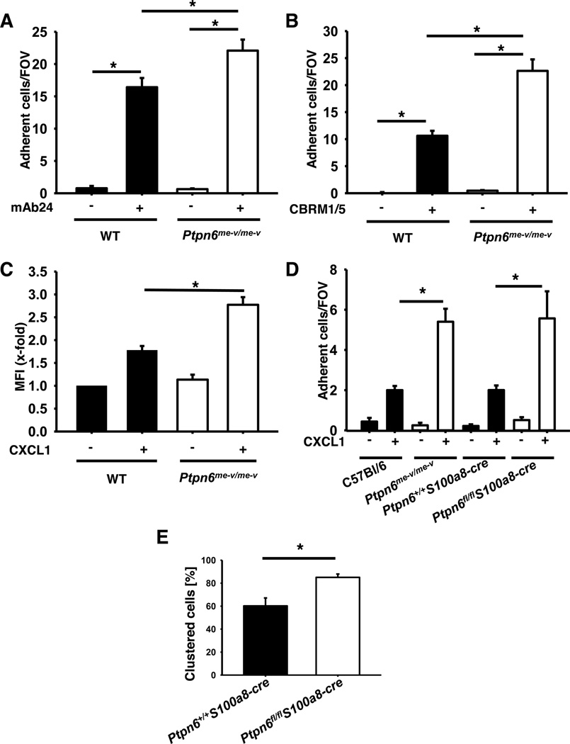
Figure 3. Gαi-signaling is regulated by Shp1.
(A–B) The number of adherent HL-60 cells (scrambled and construct 2) on a flow chamber coated with G protein, P-selectin, IL-8 and a control IgG antibody or reporter antibodies for LFA-1 (A) or Mac-1 (B) activation. (C) Adherent cells per field of view were counted and means ± SEM are displayed. ICAM-1 binding of unstimulated or CXCL1 stimulated (100 ng/ml, 3 min, 37°C) neutrophils from wild type and Ptpn6−/− mice. ICAM-1 binding was measured by FACS. (D) Number of adherent cells in wild type and Ptpn6−/− mice on capillaries coated with P-selectin or P-selectin in combination with CXCL1. Percentage of rolling neutrophils showing LFA-1 clustering in postcapillary venules of the cremaster muscle of wild type and Ptpn6−/− mice 2 h following intrascrotal TNF-α injection (E). * p < 0.05.

