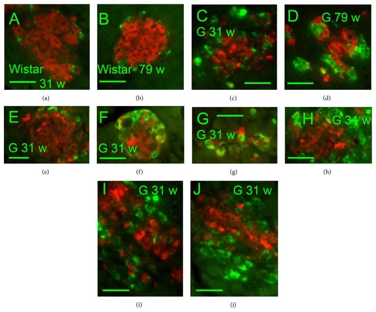
Figure 1.
Insulin and somatostatin immunohistochemical pattern of pancreatic islets. Insulin: red, SST: green, and “w”: weeks. Overlay images of pancreatic islets of 31- and 79-week-old Wistar (a, b) and Goto-Kakizaki (c–j) rats “G.” Individual or a few SST-positive delta cells were observed in Wistar controls of both ages (31 and 79 weeks) (a, b). In GK rats, increased number of strongly positive delta cells containing SST was recorded predominantly in the PI periphery as well as strongly irregularly in both ages of 31 (c, e–j) and 79 weeks (d). Original magnification 400x; scale bars 50 μm.

