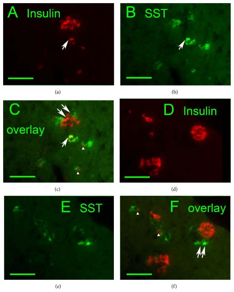
Figure 2.
Delta cell proliferation in exocrine glandular tissue ((a), (d) insulin: red, (b), (e) SST: green, and (c), (f) overlays) of 79-week-old Goto-Kakizaki rats. Individual exocrine cells present simultaneous content of insulin and SST (arrows). Coupling of individual SST- and insulin-positive endocrine cells was frequently noticed (arrowheads). Image of small proliferating endocrine masses indicates also beta and delta cells polarization (double arrows). Original magnification 400x; scale bars 50 μm.

