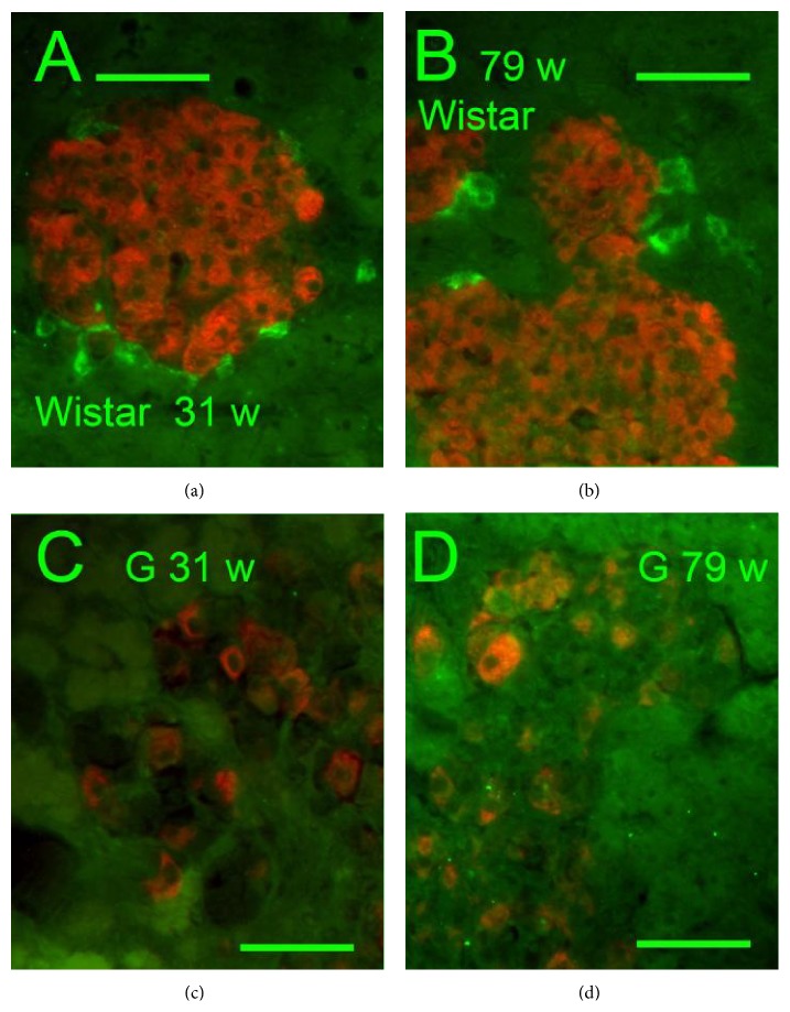
Figure 7.
Insulin and PPY immunohistochemical pattern of islets. Insulin: red, PPY: green, and “w”: weeks. Overlay images of PI of Wistar (a, b) and Goto-Kakizaki rats (c, d). Common pattern of PPY positive cells was observed in Wistar controls of both ages of 31 (a) and 79 weeks (b). In GK, no PPY positive cells were seen in both ages of 31 (c) and 79 weeks (d). Original magnification 600x; scale bars 50 μm.

