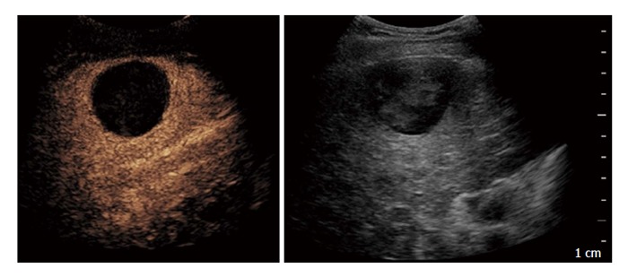
Figure 1.

Contrast-enhanced ultrasound performed after 1 mo in a 71-year-old man treated with trans-catheter arterial chemo-embolisation: On the left side complete necrosis is depicted as an avascular area; on the right side B-mode imaging of the treated area.
