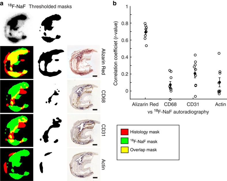
Figure 2. 18F-NaF uptake correlates with calcification but none of the histological inflammatory markers.
(a) Representative images of 18F-NaF autoradiography signal overlap with IHC-stained sequential sections. Green: 18F-NaF signal, red: histology signal, yellow: overlap. Scale bar, 1 mm. (b) High correlation is observed between 18F-NaF and Alizarin Red calcification staining, while low correlation is seen between 18F-NaF autoradiography and inflammatory marker IHC signals.

