
Fig. 3.
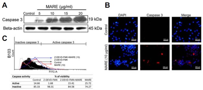
(A) MARE-treated cells were lysed and subjected to 15% SDS-PAGE. Caspase-3 protein was detected by western blotting. β-actin was used a loading control. (B) Cells were grown on cover-slips in 24-well plates. Cells were treated with 0 (control) and 10 μg/ml of MARE for 24 h. After treatment, cells were fixed with 4% PFA and were incubated overnight with the primary antibody (cleaved caspase-3) at 4°C. Cells were stained with a secondary antibody and observed under fluoresce microscope (200X). Results indicated there is an increase in the activity of caspase 3 in the cells’ nucleus (C).The activity of caspase-3 in MARE-treated cells was evaluated by flow cytometry using a caspase-3 antibody by indirect staining and the results indicate that, upon co-treatment of caspase-3 inhibitor (Z-DEVD-FMK) with MARE, the ability of MARE to activate caspase-3 is reduced.