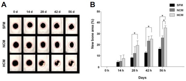
Fig. 4.

CT scanning of bone remodeling after injection of CM into a calvarial bone defect site in vivo. (A) CT images were taken at 0, 14, 28, 42, and 56 days. (B) The area of regenerated bone in the HCM treatment group was significantly greater than that of the SFM and NCM treatment groups. Data are expressed as the mean ± SD, *P < 0.05.
