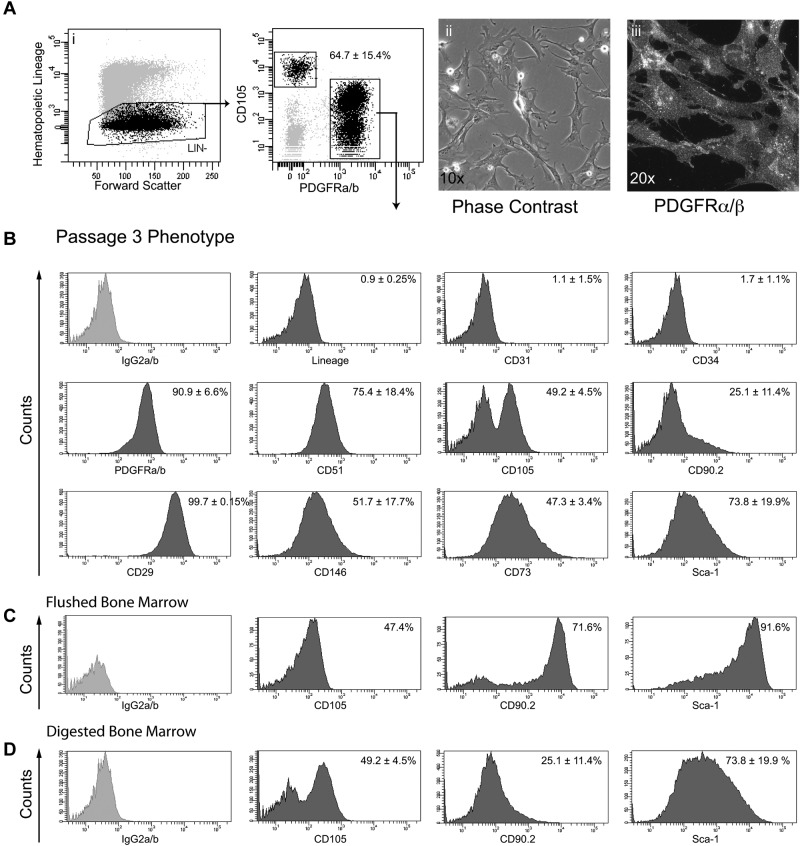
Figure 4.
Isolation and phenotypic analysis of long-term cultured Lin−PDGFRαβ+ BMSCs. (Ai-ii) Representative gating strategy of viable cells for FACS isolation of Lin−PDGFRαβ+ cells from P0 cultures. (Aii-iii) Phase-contrast image and PDGFRβ immunostaining at passage 3. (B) FACS analysis of MSC markers in cultures of passage 3 Lin−PDGFRαβ+ cells (n = 3). FACS analysis demonstrating phenotypic differences between flushed BM (C) and DBM cells (D). FACS data were collected on BD LSR II, and postacquisition analysis was performed with BD FACS Diva Version 6.1.3. Data are mean ± SD.

